Digital X-Rays & Advanced Diagnosis
Precise, low-radiation digital imaging for accurate diagnosis and better treatment planning.
Digital X-Rays & Advanced Diagnosis
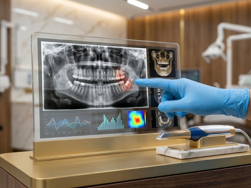
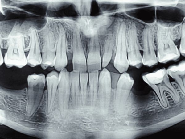
We use state-of-the-art digital radiography and cone beam computed tomography (CBCT) to produce detailed, high-resolution images of teeth, bone, nerves, and surrounding structures — instantly and with up to 90% less radiation than traditional film X-rays. Digital images are displayed chairside in seconds, allowing your dentist to discuss findings with you in real time.
Our 3D CBCT scanner provides a full volumetric view of the jaws and skull, which is invaluable for implant planning, assessing impacted teeth, diagnosing complex root canal anatomy, evaluating bone volume before surgery, and detecting pathologies that 2D X-rays may miss. This technology leads directly to better-planned, safer, and more predictable treatments.
We also use intraoral cameras to capture close-up images of individual teeth and soft tissues. These images are shown to you on screen, helping you understand your diagnosis and treatment options clearly. Transparency is central to our approach — we believe every patient should fully understand their oral health before agreeing to any treatment.
What to Expect
A fast, comfortable imaging session that informs every treatment decision.
Assessment
Your dentist determines which imaging modality is needed based on your symptoms, treatment plan, or routine check-up requirements — ensuring you receive only the imaging necessary.
Imaging
Digital X-rays or a CBCT scan are taken in a matter of seconds. The process is completely painless — you simply bite on a small sensor or stand in position for the scanner. Images appear on screen immediately.
Review & Explanation
Your dentist reviews the images with you chairside, explaining exactly what they show in plain language. Intraoral camera images may also be shared to give you a complete picture of your dental health.
Treatment Planning
Findings from imaging directly inform a personalised treatment plan. For complex cases, digital data is imported into planning software to design implant placements, surgical guides, or orthodontic setups with precision.
Why Choose This Treatment
3D Treatment Planning
CBCT scans provide a complete volumetric model of your anatomy, enabling precise implant placement, surgical planning, and orthodontic analysis.
Ultra-Low Radiation
Digital sensors require up to 90% less radiation than conventional film X-rays, making imaging safer for patients of all ages including children.
Instant Results
Images are available chairside within seconds — no waiting for film development. Your dentist can diagnose and begin planning treatment in the same appointment.
Early Problem Detection
High-resolution imaging detects decay between teeth, bone loss, cysts, and root problems at an early stage when treatment is simpler and less costly.
Before & After
Real results from real patients — images provided by the clinic.
 Before
 After
After
Common Questions About Digital Diagnosis
Yes. Our digital X-ray systems use up to 90% less radiation than traditional film X-rays, and the dose from a full-mouth series is comparable to the background radiation received during a short flight. We follow ALARA principles (As Low As Reasonably Achievable), taking only the images clinically necessary and using lead aprons for protection.
A Cone Beam CT (CBCT) scan is a 3D imaging technique that produces a full volumetric model of your teeth, jaws, nerves, and bone in a single 15-second scan. It is used when planning dental implants, assessing impacted wisdom teeth, diagnosing complex root canal anatomy, or evaluating jaw bone quality. Not every patient needs a CBCT — your dentist will advise when one is necessary.
The frequency depends on your individual oral health. Patients with a low risk of decay and gum disease may need X-rays every 18–24 months, while those with active issues or complex restorations may need them more frequently. We follow evidence-based guidelines and only take X-rays when they will meaningfully inform your care — never routinely.
Absolutely. We display all images chairside and walk you through exactly what they show. You are also welcome to request a copy of your digital images for your personal records or for use with another dental provider. Transparency is central to how we work — we believe informed patients make better decisions about their health.
Ready to Book Your Appointment?
Our specialists are ready to assist you. Get in touch today.